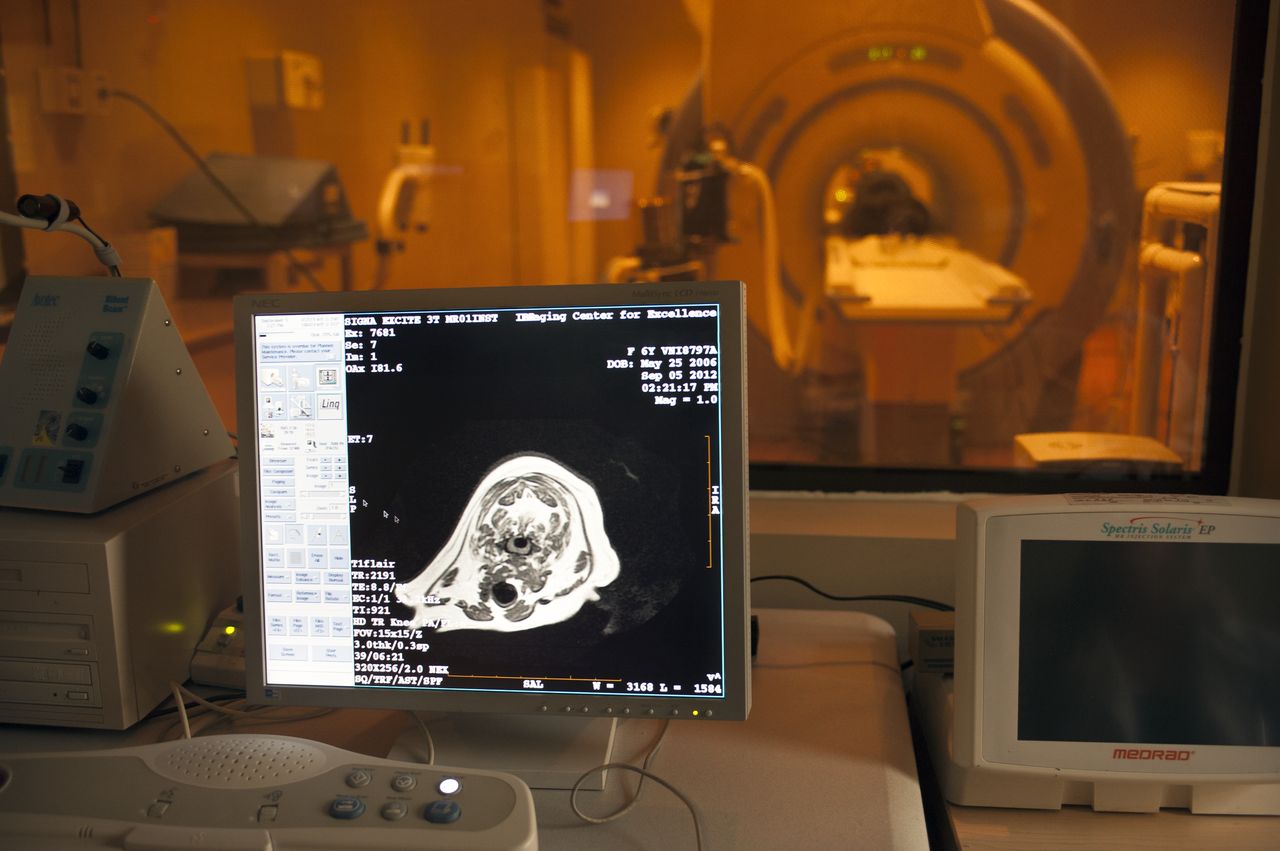
Four-legged patients to benefit from imaging grant to MSU
Contact: Karen Templeton

Photo by: Megan Bean
STARKVILLE, Miss.--Animal imaging at Mississippi State's College of Veterinary Medicine is helping advance pet health, and a new grant should take the technology even further.
Dr. Alison Plumley, a veterinary resident at the university, recently received funding from American College of Veterinary Radiology to conduct research in advanced magnetic resonance imaging techniques in dogs.
Plumley's award is the first for this specific type of imaging study provided by the non-profit organization based in Harrisburg, Pennsylvania. For more, visit www.acvr.org.
A graduate of Washington State University's College of Veterinary Medicine, she came to MSU last year through a residency match program.
Plumley said MSU's veterinary radiology program was highly recommended and carries a strong reputation. "Being here gives me ample opportunity to research novel imaging techniques," she said.
She and her mentor Dr. Jennifer Gambino, a CVM assistant professor, will use non-invasive imaging to examine different chemicals in dogs' brains to assess their normal biochemical profiles. In the future, information they uncover hopefully will help better diagnose inflammation, infection, tumors and other brain diseases.
"Normally, animal and human patients have to undergo brain biopsies to get information on a tumor," Plumley explained. "These procedures are timely, invasive and sometimes can present further risk. Imaging can assist neurologists in obtaining the information needed to determine the best treatment plans."
Their research and imaging work primarily will take place at the Veterinary Specialty Center, an affiliate of the CVM, and MSU's Institute for Imaging and Analytical Technologies.
Using imaging technology to understand more about canine brain tumors, the center is part of Starkville's Premier Imaging complex located at the intersection of Stark Road and State Highway 182. Premier treats human patients, while VSC treats animal patients.
As the first of a two-part process, Plumley, Gambino and other team members will be comparing results from two different types of magnetic resonance spectroscopies--advanced single and multivoxel--to quantify the molecular brain chemicals.
Single voxel imaging's advantages include a capability to be run quickly and provide detailed information. Multivoxel imaging is more robust and specific, yielding pages of data that can require more time for clinical interpretation.
A board-certified veterinary radiologist, Gambino said a single voxel can be run "in seven-to-eight minutes," adding, "In people, research has shown these techniques to be effective methods of obtaining information on brain tumors, infections and inflammatory disease states."
She continued: "Multivoxel will give us specifics that can take a lot more time to run and decipher. It is to our advantage to see what viable information we can gain from shorter processes, so we can gain the greatest amount of detail with the least invasive of techniques to guide further diagnostics and treatments."
Imaging increasingly has become an important tool in research and clinical care. Current technologies can provide incredible details about the brain and brain-tumor structures, even including functional data down to the molecular level.
"The goal is to eventually identify different types of tumors based on the combined results of the conventional MR images and MRS so we can better provide recommendations on treatment, whether surgery, radiation, chemotherapy or a combination thereof," Gambino said. "With the plethora of information gleaned from the imaging, we can make effective treatment decisions before approaching any type of invasive techniques. This is something that also has a huge impact on human health."
The second part of the team's investigation will seek to determine whether lower doses of a solution called gadolinium can be effective for diagnostic imaging.
Plumley said gadolinium "is used safely in humans and animals all of the time and provides additional information with regard to brain disease or lack thereof." As with other contrast agents or drug therapies, however, "reactions can occur," she noted.
"Currently, the dose of these agents in dogs and cats is extrapolated from the use of the agents in people," she said. "Evidenced-based dosing is very desirable to minimize any such deleterious side effect, despite them being rarely seen in practice. Our main goal is to improve safety while maximizing benefits seen when using contrast and improving the diagnostic quality of our MR studies."
Second-year veterinary student Emerald D. Barrett of Louisville will be spending the next five months analyzing results of the team's research and publishing the findings. A 2014 magna cum laude MSU graduate in animal and dairy science/pre-veterinary medicine, she now is enrolled in the college's Summer Research Experience Program.
"Students are the nuts and bolts of these kinds of projects," Gambino said of Barrett and others. "We really depend on their extra set of eyes and perspective."
This is one of the few places in the country with the type of equipment, specialized software and trained personal to do this kind of research. It's a great place to be."
MSU, Mississippi's flagship research university, is online at www.msstate.edu, facebook.com/msstate, instagram.com/msstate and twitter.com/msstate.